Knee Muscle Anatomy Mri - Atlas of Knee MRI Anatomy - W-Radiology : Free access interactive and dynamic anatomical atlas.
Knee Muscle Anatomy Mri - Atlas of Knee MRI Anatomy - W-Radiology : Free access interactive and dynamic anatomical atlas.. Click now to learn more about the bones, muscles, and soft tissues of these regions at leg and knee anatomy: See the pictures and anatomy description of knee joint bones, cartilage, ligaments, muscle and tendons with resources for knee problems & injuries. This mri knee cross sectional anatomy tool is absolutely free to use. 4, infrapatellar fat pad of hoffa. Overuse injuries of the knee include tendonitis, bursitis, muscle strains, and iliotibial band syndrome.
Magnetic resonance imaging (mri scan): An understanding of normal anatomy and biomechanics of the knee extensor mechanism is necessary to comprehend the imaging of extensor mechanism injuries. Use the checklist to quiz yourself. Current and accurate information for patients about magnetic resonance imaging (mri)of the knee. Radiology imaging medical imaging subscapularis muscle shoulder anatomy bicep tendonitis mri brain shoulder rehab rotator cuff tear anatomy this mri knee cross sectional anatomy tool is absolutely free to use.
Leg Mri Anatomy - soupbranch.com from radsource.us The muscles of the knee include the quadriceps, hamstrings, and the muscles of the calf. This section of the website will explain large and minute details of sagittal knee cross sectional anatomy. The journal of musculoskeletal medicine. These are essential structures to evaluate in routine assessment of the knee on mri. Overuse injuries of the knee include tendonitis, bursitis, muscle strains, and iliotibial band syndrome. This mri knee cross sectional anatomy tool is absolutely free to use. Song, uc san francisco msiv gillian lieberman md. It is a noninvasive test that can visualize the inner structures of the knee, including the cartilage and ligaments, the surface of the bones, and the muscles and tendons that surround the knee joint.
Rubin da, kettering jm, towers jd, britton ca:
There are various muscles that control movement, ligaments that. Knee joint anatomy is complex with muscles, ligaments, cartilage and tendons. Click now to learn more about the bones, muscles, and soft tissues of these regions at leg and knee anatomy: An exercise program can strengthen the muscles surrounding the knee, increasing the knee's stability. The journal of musculoskeletal medicine. Want to learn more about it? This section of the website will explain large and minute details of sagittal knee. Learn about mri anatomy with free interactive flashcards. Current and accurate information for patients about magnetic resonance imaging (mri)of the knee. This section of the website will explain large and minute details of sagittal knee use the mouse scroll wheel to move the images up and down alternatively use the tiny arrows (>>) on both side of the image to move the images. See the pictures and anatomy description of knee joint bones, cartilage, ligaments, muscle and tendons with resources for knee problems & injuries. Knee anatomy francesc malagelada jordi vega pau golanó the knee is the largest joint in the human body and one of the most complex from a functional point of view. Mr imaging of knees having isolated and combined ligament injuries.
Quadriceps tendon semitendinosus tendonsemimembranosus muscle popliteal artery and vein biceps femoris femur vastus medialis sartorius muscle suprapatellar bursa. If you think of the knee in layers, the deepest layer is bone and ligaments, then ligaments of the joint capsule, then muscles on top. Scroll through the structures to understand the anatomy. This webpage presents the anatomical structures found on knee mri. Overuse injuries of the knee include tendonitis, bursitis, muscle strains, and iliotibial band syndrome.
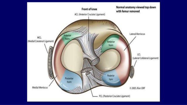
Mri anatomy of knee Dr. Muhammad Bin Zulfiqar from image.slidesharecdn.com Current and accurate information for patients about magnetic resonance imaging (mri)of the knee. This webpage presents the anatomical structures found on knee mri. Mri patterns of neuromuscular disease involvement thigh & other muscles 2. Anatomy of peritoneum and mesentery. Learn anatomy using a full pacs! Knee anatomy francesc malagelada jordi vega pau golanó the knee is the largest joint in the human body and one of the most complex from a functional point of view. This mri knee cross sectional anatomy tool is absolutely free to use. Mr imaging of knees having isolated and combined ligament injuries.
Injuries of the patellofemoral joint.
Song, uc san francisco msiv gillian lieberman md. Each anatomical structure was labeled interactively. Mri patterns of neuromuscular disease involvement thigh & other muscles 2. If you think of the knee in layers, the deepest layer is bone and ligaments, then ligaments of the joint capsule, then muscles on top. General anatomy and musculoskeletal system. Rubin da, kettering jm, towers jd, britton ca: Single mri images are called slices. Magnetic resonance imaging (mri) is the test of choice to confirm the diagnosis of a torn meniscus. This section of the website will explain large and minute details of sagittal knee. Free access interactive and dynamic anatomical atlas. It is a noninvasive test that can visualize the inner structures of the knee, including the cartilage and ligaments, the surface of the bones, and the muscles and tendons that surround the knee joint. Want to learn more about it? Learn about mri anatomy with free interactive flashcards.
Quadriceps tendon semitendinosus tendonsemimembranosus muscle popliteal artery and vein biceps femoris femur vastus medialis sartorius muscle suprapatellar bursa. General anatomy and musculoskeletal system. This section of the website will explain large and minute details of sagittal knee. See the pictures and anatomy description of knee joint bones, cartilage, ligaments, muscle and tendons with resources for knee problems & injuries. Magnetic resonance imaging (mri) is the test of choice to confirm the diagnosis of a torn meniscus.
Shoulder: MRI, radiographical, and illustrated anatomical ... from www.imaios.com Magnetic resonance imaging (mri scan): 1 november 2002 mri anatomy of the knee and shoulder james y. Each anatomical structure was labeled interactively. Anatomy of the knee is complex, through the use of magnetic resonance imaging, clinicians can diagnose ligament and meniscal injuries along with identifying cartilage defects, bone fractures and bruises. Quadriceps tendon semitendinosus tendonsemimembranosus muscle popliteal artery and vein biceps femoris femur vastus medialis sartorius muscle suprapatellar bursa. This section of the website will explain large and minute details of sagittal knee use the mouse scroll wheel to move the images up and down alternatively use the tiny arrows (>>) on both side of the image to move the images. Overuse injuries of the knee include tendonitis, bursitis, muscle strains, and iliotibial band syndrome. Knee anatomy francesc malagelada jordi vega pau golanó the knee is the largest joint in the human body and one of the most complex from a functional point of view.
This webpage presents the anatomical structures found on knee mri. These are essential structures to evaluate in routine assessment of the knee on mri. 12 photos of the knee muscle anatomy mri. Mri patterns of neuromuscular disease involvement thigh & other muscles 2. Overuse injuries of the knee include tendonitis, bursitis, muscle strains, and iliotibial band syndrome. An understanding of normal anatomy and biomechanics of the knee extensor mechanism is necessary to comprehend the imaging of extensor mechanism injuries. See the pictures and anatomy description of knee joint bones, cartilage, ligaments, muscle and tendons with resources for knee problems & injuries. A knee mri (magnetic resonance imaging) scan uses energy from strong magnets to create pictures of the knee joint and muscles and tissues. Scroll through the structures to understand the anatomy. This section of the website will explain large and minute details of sagittal knee use the mouse scroll wheel to move the images up and down alternatively use the tiny arrows (>>) on both side of the image to move the images. Knee anatomy francesc malagelada jordi vega pau golanó the knee is the largest joint in the human body and one of the most complex from a functional point of view. There are various muscles that control movement, ligaments that. General anatomy and musculoskeletal system.